
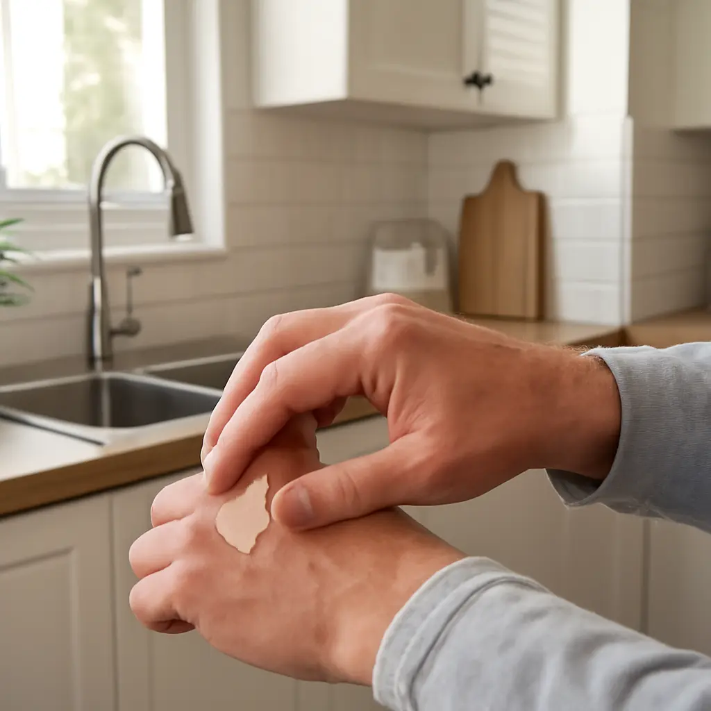
Hand skin heals differently from skin on many other parts of the body because it is repeatedly exposed to friction, washing, chemicals, and movement—and because the palm and back of the hand have different structures. That matters when removing a splinter, treating a cut, or deciding whether a wound needs professional care: a small injury over a finger joint may be more consequential than its size suggests. The American Academy of Dermatology recommends keeping minor wounds clean and moist with petroleum jelly and a dressing, while hand-surgery guidance emphasizes prompt assessment for deep, contaminated, or function-limiting injuries. Understanding hand skin anatomy, healing conditions, and warning signs helps match removal and wound care to the actual risk.
Hand Skin Healing: Anatomy and Recovery Differences
Hand skin healing is the repair of the skin barrier and underlying tissue after injury, influenced by the hand’s specialized anatomy, blood supply, motion, and frequent contact with irritants. The National Library of Medicine’s StatPearls overview of wound healing describes the process in overlapping stages: hemostasis, inflammation, tissue growth, and remodeling. The sequence is broadly shared across the body, but hand wounds face practical stresses that can disrupt it, including repeated bending, pressure, moisture, and contamination.
The distinction is not that every hand wound heals slowly. Rather, the location and demands of the hand can make certain injuries harder to protect and more likely to affect movement or sensation. The American Society for Surgery of the Hand notes that apparently small hand injuries can involve tendons, nerves, blood vessels, joints, or bones. That is why removal decisions should begin with what is embedded, where it is, and whether normal hand function has changed.
Palm skin and fingertip skin
Palmar skin is thick, hairless, and adapted to gripping and repeated pressure. Fingertips also contain dense sensory structures that support fine touch. A shallow scrape on the palm may be less concerning than a deep puncture, but a wound near a fingertip can be painful and may affect sensation or precision. Avoid digging into either area to remove a deeply lodged object: probing can enlarge the wound or injure structures beneath the skin.
Dorsal hand skin and joints
The skin on the back of the hand is generally thinner and more mobile than palmar skin, with less protective padding over some underlying structures. Cuts over knuckles or finger joints are exposed to repeated stretching whenever the hand bends. A wound that crosses a joint, gapes when the finger moves, or limits motion warrants prompt medical assessment; clinicians may need to check tendon function, joint involvement, and whether closure is appropriate.
Healing stages and hand-specific stresses
Wound healing starts with clotting, followed by inflammation, new tissue formation, and remodeling. These stages overlap and do not follow an identical timetable for every person or injury. On the hand, frequent washing and work can repeatedly expose a wound to water, friction, or chemicals. The Centers for Disease Control and Prevention also identifies diabetes and immune compromise among factors that increase infection risk or complicate recovery, making individualized medical advice especially important.
Removal Decisions: Match the Method to the Hand Injury
Removal means taking out an object or damaged tissue only when doing so is safe and appropriate. A visible, superficial splinter is different from a fishhook, glass fragment, deeply embedded thorn, or object near a joint, nail, tendon, or nerve. The American Academy of Dermatology advises cleaning minor wounds and protecting them with petroleum jelly and a bandage; it does not make home exploration a safe substitute for clinical evaluation when an object is deep or difficult to grasp.
The practical rule is to avoid making a small wound larger in pursuit of an object. Remove only what is clearly superficial and readily accessible, and seek care if it is deep, painful, contaminated, or associated with altered function or sensation. These categories connect the type of object to the risks created by the hand’s anatomy.
Superficial splinters
A superficial splinter is a small fragment near the skin surface that can be grasped without cutting or digging. Wash your hands and the area with soap and water. If the end is plainly visible, clean tweezers may be used to draw it out along its entry path. Stop if it breaks, resists, or causes substantial pain; repeated attempts can push fragments deeper or damage surrounding tissue. After removal, rinse the area, apply a thin layer of petroleum jelly, and cover it with a clean dressing.
Deep, contaminated, or high-risk objects
A deep splinter, glass shard, fishhook, or object embedded near a joint, nail bed, or tendon should be assessed by a clinician rather than extracted by force at home. Do not squeeze or probe a puncture wound. The Mayo Clinic’s first-aid guidance advises medical attention for deep punctures, wounds containing debris that cannot be removed, and injuries with signs of infection. A clinician can decide whether imaging, local anesthesia, specialized removal, wound closure, or tetanus protection is needed.
Cuts, torn skin, and wound closure
For a minor cut, apply steady pressure with clean material, rinse away visible dirt with clean running water, and cover it with a clean dressing. Do not pour harsh antiseptics into the wound, as they can irritate tissue. A deep or gaping cut, one that will not stop bleeding, or one that exposes deeper tissue needs urgent evaluation. Hand wounds may require a clinician to check movement, sensation, and circulation before deciding whether stitches or another form of closure is suitable.
Aftercare: Protect the Barrier and Preserve Function
Aftercare supports the skin barrier while avoiding unnecessary restriction of hand movement. The American Academy of Dermatology recommends keeping minor wounds moist with petroleum jelly and covered, changing the dressing daily or when it becomes wet or dirty. A clean, protected wound is less exposed to friction and contamination, but dressings should not be so tight that fingers become numb, pale, cold, or increasingly painful.
Cleaning and dressing changes
Gently rinse a minor wound with clean water and mild soap around the area. Reapply a thin layer of petroleum jelly and use a suitable clean bandage. If a dressing sticks, moisten it before removal rather than pulling at healing tissue. People whose work involves food, caregiving, tools, or chemicals may need more frequent dressing checks and should follow workplace hygiene rules without allowing the wound to remain wet or soiled.
Movement, friction, and return to activity
Repeated gripping and bending can pull at wounds, particularly those on knuckles or across creases. Protect the injured area during activities that could reopen or contaminate it. At the same time, do not immobilize a finger or hand for longer than advised: stiffness can become a problem, and some injuries need guided movement or specialist follow-up. Follow the treating clinician’s instructions if the wound is deep, closed with stitches, or involves a joint or tendon.
Infection warning signs and tetanus review
Seek medical advice for spreading redness, increasing warmth or swelling, worsening pain, pus, fever, red streaks, or reduced movement. Prompt care is also appropriate for numbness, tingling, pale or blue fingers, uncontrolled bleeding, or a wound that appears deep. The CDC recommends reviewing tetanus vaccination for wounds; whether a booster is needed depends on wound type and vaccination history. A clinician or local public-health guidance can determine the appropriate timing, especially after dirty or puncture wounds.
Real-World Risk: Why Injury Location Changes the Plan
Consider two injuries caused by a wood splinter: one fragment lies just beneath the surface of the palm and is easily grasped; another is deeply embedded beside a finger joint and hurts when the finger bends. The first may be appropriate for cautious home removal and routine wound care. The second merits professional assessment because the location and movement-related pain raise concerns beyond the visible fragment. The same principle applies to cuts: location, depth, contamination, and function are more informative than wound size alone.
For a visual aid, an article or clinic handout could compare a palm, fingertip, and knuckle wound, labeling nearby joints, tendons, and sensory areas. Such a diagram should support—not replace—clinical evaluation, because surface appearance cannot reliably show the full depth or path of an object.
Conclusion: Let Hand Anatomy Guide Removal and Recovery
Hand skin healing follows the body’s familiar repair stages, but palm and fingertip structure, thinner skin over the back of the hand, constant movement, and frequent exposure change how an injury should be managed. A clearly superficial splinter may be removed gently; deep, contaminated, or function-affecting injuries should not be explored at home. Clean, moist wound care and suitable protection support minor wounds, while worsening symptoms, altered sensation, restricted motion, or tetanus concerns call for medical advice. Choose care based on the object and its location, not just the apparent size of the wound, and consult a health professional when in doubt.
Sources: American Academy of Dermatology, “How to Treat Minor Cuts,” https://www.aad.org/public/everyday-care/injured-skin/burns/treat-minor-cuts; American Society for Surgery of the Hand, “Common Hand Injuries,” https://www.assh.org/handcare/condition/common-hand-injuries; National Library of Medicine, StatPearls, “Wound Healing Phases,” https://www.ncbi.nlm.nih.gov/books/; Centers for Disease Control and Prevention, “Clinical Guidance for Wound Management to Prevent Tetanus,” https://www.cdc.gov/tetanus/hcp/clinical-guidance/index.html; Mayo Clinic, “Puncture Wounds: First Aid,” https://www.mayoclinic.org/first-aid/first-aid-puncture-wounds/basics/art-20056665.